
A)The Gram Stain:
CAUTIONS in Procedure:
(1) Don't make your smear too thick or the theme of the Gram stain is ruined... thick = purple even if you have a Gram negative (pink) as the entire stain detects the "thickness" of the cell wall and 100 thin cells on top of each other = thick or purple!
(2) USE 80% ethanol, alcohol, or acetone alcohol NOT ACID ALCOHOL!
CAUTIONS in READING the gram stain:
(Slide A#1) Sometimes when staining the Bacillus there are old cells in the culture. Old cells "fall apart" and become "leaky" so that they stain pink or Gram negative when they should stain Gram positive (purple)... READ only the cells ON TOP (foreground) of the stain, not those pink cells that have fallen apart and are underneath the purple rods.
(Slide A#2) Mycobacterium smegmatis is a problem organism on all 3 major stains... on the Gram stain it stains Gram variable on the positive side (80% positive or Purple with a few Pink rods) but it has a thin peptidoglycan layer but stains positive because of the thick lipid layer which "traps" Crystal Violet and thus gives the vegetative cell the purple Gram positive appearance at 1000x (3) The Gram staining is read as "the majority color" wins.. except when there are a majority of old fallen apart pink dead cells underneath large intact PURPLE rods = Pos
EXAMPLE PHOTOS:
Slide A#1: Gram Positive (purple) rods ON TOP with old dead pink cells underneath =GRAM POS
Slide A#1: Gram Positive (purple) rods ON TOP with old dead pink cells underneath =GRAM POS
  |
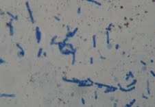
Slide A#2 Gram variable on the positive side rods (Mycobacterium)
|
B) THE ACID FAST STAIN:
CAUTIONS in Procedure: (1) When staining the Acid Fast Mycobacterium smegmatis note that the culture grows on a slant with a "scabby" appearance while the grow on your loop is "sticky/gooey" and thus difficult to smear... try not to get too much while "breaking-up" any clumping as much as you can. (2) USE the ACID ALCOHOL NOT ethanol or acetone alcohol... wash off the primary stain lightly after heating it in a flame and after the Acid Alcohol step.
CAUTIONS in READING the stain: (Slides B#1 & B#2) This stain is read differently than any other... YOU MUST MOVE THE SLIDE AROUND THE CIRCLE AFTER FOCUSING (it is quicker to scan on 10X and to put any hot pink areas at the end of the pointer THEN go to 100X). You aren checking the entire circle for hot pink or fuchsia rods. ONE fuchsia rod (Slide B#1) makes the stain positive or ACID FAST. The majority of the stained rods could be baby blue and one fuchsia rod would make the stain positive or all hot pink (Slide B#2) and a few baby blue. A properly "cooked AF stain looks like Slide B#3. Slide #4 where there are all baby blue cells is AF negative or Non-acid Fast. (Slide B#4). Any Bacillus ??? ENDOSPORE might take-up the fuchsia stain... but!!!! endospores are NOT cells and they are NOT RODS... they are oval or round (Slide B#5)!
     | |
| Slide B#5 is an Acid Fast stain containing Bacillus megaterium (Non-acid fast baby-blue "hot-dog shaped" rods and hot pink oval repeating endospores... which are NOT CELLS! |
C) THE ENDOSPORE STAIN:
CAUTIONS in Procedure: (1) Only the rod-shaped genera Bacillus (aerobic) and Clostridium (anaerobic) produce resistant survival structures known as ENDOSPORES. (2) DO NOT USE ANY ALCOHOL on this stain... there are only 3 liquids: to the smear add a few drops of Malachite green, heat through a flame 5x, wash lightly with water, apply Safranin for 4 minutes, wash, blot and view under 1000x.
CAUTIONS in READING the stain (the most difficult stain to read):
(Slide C#1) Enspores may appear as green spheres/ovals or colorless and rarely as small round structure that appear to be clear holes in pink rods... REMEMBER there must be pink vegetative rods involved as Bacillus and Clostridium!
*Endospores are always oval (egg-shaped) or round and smaller than the pink vegetative cell that made them...
*Endospores are NEVER SHAPED LIKE RODS
*ENDOSPORES are never larger than the pink vegetative cells. Pink rods with green ovals inside or extruded nearby as green ovals or spheres are read as Endospore positive. Pink rods with empty holes are read as ENDOSPORE postive.
*YOUR LAST AND NEVER FIRST thought is that when you see All green ovals... it is read as Endospore positive.
*REMEMBER ALL GREEN (unless they are all and only green ovals - endospore) ON ENDOSPORE OR ALL HOT PINK ON ACID FAST IS ALWAYS WRONG OR OVERCOOKED!
(Slide C#4) Pink and green rods of the same size or with various size green objects are read as Endospore negative as various-sized green things are cell garbage. All pink vegetative cells are read as Endospore negative.
CAUTION:!!! The lipid layer of theMycobacteria smegmatis (Slide C#3) may "take-up" the Malachite green and appear green. This microbe under Endospore staining may present as pink and green rods of the same size or green rods with smaller red rods... this is NOT oval or round endopores and is thus read as Endospore negative.
    | |
| Endospore positive stain.... pink rods with empty holes AND green oval endospores |